
Khalifa University builds AI cancer scan model
New model beats rivals across 13 tests, mimics how pathologists actually analyse

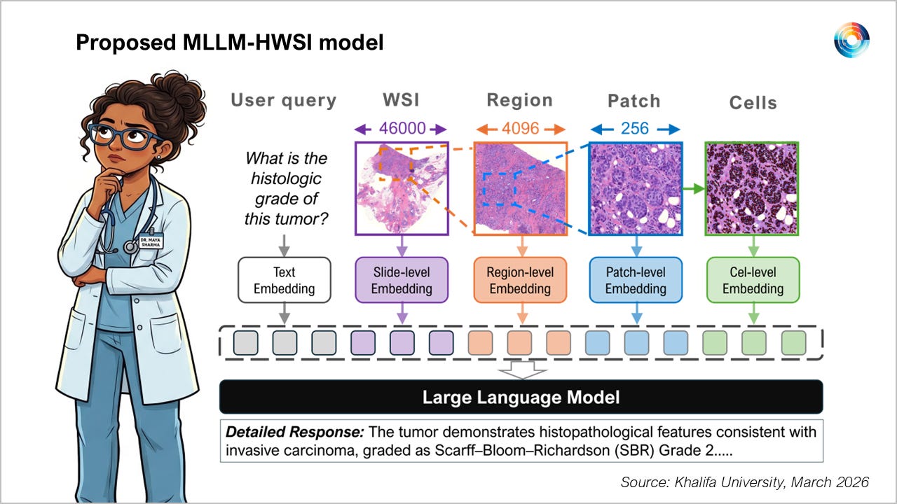
#UAE #healthcare - Researchers from Abu Dhabi research university Khalifa University have presented a new AI model built to read cancer pathology slides like human pathologists do at CVPR 2026 in Colorado earlier this month, Most existing AI models compress an entire tissue slide into a single data summary, losing fine detail in the process. Khalifa University’s MLLM-HWSI model reads slides at four levels at once, from individual cells through to the whole slide, mirroring how pathologists actually move between zoomed-in and zoomed-out views. The model outperformed AI models relying on prior approaches across 13 public benchmarks, covering six diagnostic tasks.
SO WHAT? - Cancer diagnosis from tissue slides still relies on a pathologist scanning a gigapixel image by eye across multiple magnifications, holding cellular detail and overall tissue structure in mind at once. Most AI tools built to scan slides for cancer indications simply summarise the whole image, which loses the fine-grained evidence an informed diagnosis depends on. By building a model that reasons cell-by-cell and region-by-region before reaching a conclusion, Khalifa University’s work points toward AI tools that don’t just classify medical image data, but show their reasoning in a way a pathologist can actually check.
KEY POINTS:
Khalifa University researchers presented MLLM-HWSI, a hierarchical AI model for understanding whole slide pathology images, at CVPR 2026, the IEEE/CVF Conference on Computer Vision and Pattern Recognition held from June 3rd to 7th in Colorado.
The AI model reads tissue slides across four scales, treating cells as if they were words, small patches as phrases, larger regions as sentences, and the whole slide as a paragraph, rather than compressing everything into one summary.
It uses a Cell-Cell Attention Fusion (scCAF) transformer to combine information from segmented cells into a compact representation for each patch of tissue.
The model jointly applies a hierarchical contrastive learning method and a cross-scale consistency check to keep meaning aligned from individual cells up to the full slide.
MLLM-HWSI achieved state-of-the-art results across 13 publicly available whole slide image benchmark tests and six computational pathology tasks, including classification, retrieval and report generation.
It was tested against 24 existing state-of-the-art models in the field and consistently outperformed them, according to the research paper.
The approach is designed to produce interpretable, evidence-grounded outputs that link specific text findings, such as abnormal cell shapes, to the exact part of the image that supports them.
Researchers say future work will extend the model beyond pathology slides into radiology, genomics and clinical records, aiming for a unified medical AI system that reasons across a patient’s full record.
Researchers on the project included: Sajid Javed,, Basit Alawode, Muaz Khalifa Al-Radi, Shahad Albastaki, Moshira Ali Abdalla and Asim Khan from Khalifa University Department of Computer Science; Arif Mahmood from Information Technology University, Pakistan; and Muhammad Bilal from King Abdulaziz University (KAU), Saudi Arabia; and Mohammed Bennamoun from University of Western Australia.
[Written and edited with the assistance of AI]
Source: Khalifa University
LINKS
MLLM-HWSI research paper (arXiv)
MLLM-HWSI code (GitHub)
Read about other AI models developed by Khalifa University:
Khalifa University built AI model tops telco leaderboard (Middle East AI News)
Khalifa University unveils breakthrough RF AI model (Middle East AI News)
KU researchers build cybersecurity AI assistant (Middle East AI News)
Telecom industry partners develop Arabic Telecom LLM (Middle East AI News)